Renal Calculi (Kidney Stones): Essential Guide for NCLEX Exam

Renal calculi, commonly known as kidney stones, are a significant topic on the NCLEX exam. Understanding their formation, symptoms, treatment, and prevention is crucial for both nursing practice and exam success. This blog post covers everything you need to know about kidney stones, along with sample NCLEX questions to help you prepare.
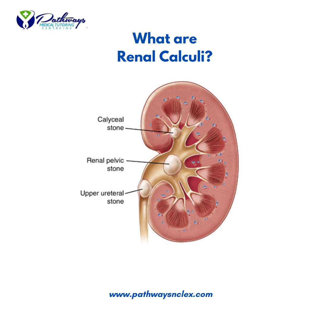
What are Renal Calculi?
Renal calculi, or kidney stones, are hard deposits of minerals and salts that form in the kidneys. They vary in size, from tiny grains to larger stones that can obstruct the urinary tract. Kidney stones can cause severe pain and complications if left untreated.
Types of Kidney Stones
- Calcium Stones: The most common type, often composed of calcium oxalate. They can also be made of calcium phosphate.
- Struvite Stones: Often form in response to an infection, such as a urinary tract infection (UTI). These stones can grow quickly and become quite large.
- Uric Acid Stones: Common in people with high protein diets, gout, or those who are dehydrated. Uric acid stones are more likely to form in acidic urine.
- Cystine Stones: Rare and usually occur in individuals with a genetic disorder called cystinuria, which causes the kidneys to excrete too much cystine.
Pathophysiology
Kidney stones form when the concentration of certain substances in the urine—such as calcium, oxalate, uric acid, or cystine—becomes so high that they crystallize and clump together. Factors that contribute to stone formation include:
- Dehydration: Lack of adequate water intake concentrates substances in the urine, increasing the risk of stone formation.
- Diet: High intake of oxalate-rich foods, excessive dietary sodium, and high-protein diets can contribute to stone formation.
- Metabolic Disorders: Conditions such as hyperparathyroidism, gout, or renal tubular acidosis can lead to stone formation.
- Urinary Tract Infections (UTIs): Certain infections can alter the chemical balance in the urine, leading to stone formation.
Risk Factors
- Family History: A history of kidney stones increases the likelihood of developing them.
- Dehydration: Inadequate fluid intake concentrates minerals in the urine, promoting stone formation.
- Dietary Factors: High intake of oxalate-rich foods, salt, and animal proteins increases the risk.
- Obesity: Excess weight can alter the acid-base balance in the body, leading to stone formation.
- Certain Medications: Some medications, such as diuretics, antacids, and certain supplements, can increase the risk.
Symptoms
Kidney stones may not cause symptoms until they move within the kidney or pass into the ureter. When symptoms occur, they may include:
- Severe Pain: Often described as sharp, cramping pain in the back and side, below the ribs, and radiating to the lower abdomen and groin (renal colic).
- Hematuria: Blood in the urine, making it pink, red, or brown.
- Nausea and Vomiting: Often occur due to the severe pain.
- Frequent Urination: An increased urge to urinate, often with little urine output.
- Dysuria: Painful urination.
- Fever and Chills: May indicate an infection, especially if a stone is obstructing urine flow.
Diagnosis
- Imaging Tests:
- CT Scan: The most common and accurate test for detecting kidney stones.
- Ultrasound: Often used for initial diagnosis, especially in pregnant women.
- X-ray: A KUB (Kidneys, Ureters, and Bladder) X-ray can show some stones, though not all types are visible.
- Urinalysis: Checks for the presence of blood, crystals, bacteria, and other abnormalities in the urine.
- Blood Tests: Assess kidney function and levels of substances that can contribute to stone formation, such as calcium and uric acid.
- Stone Analysis: If a stone is passed, it can be collected and analyzed to determine its composition.
Treatment
Conservative Management
- Increased Fluid Intake: Encouraging patients to drink plenty of water to help flush out small stones.
- Pain Management: NSAIDs or opioids may be prescribed to manage pain.
- Medications:
- Alpha Blockers: Help relax the muscles in the ureter, allowing the stone to pass more easily.
- Thiazide Diuretics: Can be prescribed to reduce calcium in the urine.
- Potassium Citrate: Used to prevent stone formation by making the urine less acidic.
Surgical Interventions
- Extracorporeal Shock Wave Lithotripsy (ESWL): Uses shock waves to break the stone into smaller pieces that can be passed in the urine.
- Ureteroscopy: A small scope is passed through the urethra and bladder into the ureter to remove the stone or break it into smaller pieces.
- Percutaneous Nephrolithotomy: A surgical procedure for removing large or complex stones directly from the kidney through a small incision in the back.
- Parathyroid Surgery: If stones are caused by overactive parathyroid glands, surgery to remove the gland may be necessary.
Prevention
- Stay Hydrated: Encourage patients to drink enough fluids to produce at least 2.5 liters of urine daily.
- Dietary Modifications:
- Reduce Salt and Protein Intake: Limit salt and animal proteins in the diet.
- Avoid Oxalate-Rich Foods: Reduce intake of foods like spinach, nuts, and tea.
- Medications: For those at high risk, medications may be prescribed to prevent stone formation.
- Regular Monitoring: Regular check-ups, especially for those with a history of kidney stones, to monitor for recurrence.
Nursing Interventions
- Pain Management: Administer prescribed analgesics and monitor the patient’s pain levels.
- Encourage Fluid Intake: Educate the patient on the importance of hydration and monitor fluid intake and output.
- Strain Urine: Collect and strain all urine to catch any stones that pass, so they can be analyzed.
- Educate the Patient: Provide information on dietary changes, medication adherence, and lifestyle modifications to prevent future stones.
- Monitor for Complications: Be alert for signs of infection, obstruction, or worsening pain, and report these to the healthcare provider.
Sample NCLEX Questions
Question 1
A patient presents with severe flank pain and hematuria. The nurse suspects a kidney stone. Which diagnostic test is most commonly used to confirm the diagnosis?
A. Ultrasound
B. X-ray
C. CT scan
D. Urinalysis
Answer: C. CT scan
Question 2
A patient with a history of recurrent kidney stones is advised to avoid foods high in oxalate. Which of the following foods should the patient limit?
A. Bananas
B. Spinach
C. Chicken
D. Milk
Answer: B. Spinach
Question 3
A nurse is caring for a patient who has just undergone extracorporeal shock wave lithotripsy (ESWL) for kidney stones. Which of the following is a priority intervention?
A. Encourage early ambulation
B. Monitor for signs of infection
C. Administer a high-protein diet
D. Restrict fluid intake
Answer: B. Monitor for signs of infection
Question 4
A patient with uric acid kidney stones is prescribed allopurinol. What is the purpose of this medication?
A. Increase calcium excretion
B. Reduce uric acid levels
C. Prevent bacterial infection
D. Alkalinize the urine
Answer: B. Reduce uric acid levels
NCLEX Preparation Tips for Renal Calculi
- Understand the Types: Be familiar with the different types of kidney stones and their causes.
- Recognize Symptoms: Know the key symptoms of renal calculi and how they differ from other conditions.
- Know the Treatments: Understand both conservative and surgical treatments for kidney stones.
- Patient Education: Be prepared to educate patients on prevention strategies, including dietary and lifestyle changes.
- Practice Questions: Use NCLEX-style questions to reinforce your knowledge and prepare for the exam.


